Brain scans could predict future risk of psychosis
By Liz Lockhart
Computer analysis of brain scans could help to predict the severity of psychosis and, by doing so, could  allow doctors to make more accurate diagnosis and treatment plans, according to a new UK study.
The use of computers to assess the risk of further of physical disease is common but no accurate tests were available for psychiatrists. Until now, brain imaging by MRIs to detect the difficult and subtle changes in the brain which are associated with psychosis have been of small benefit for clinical use.
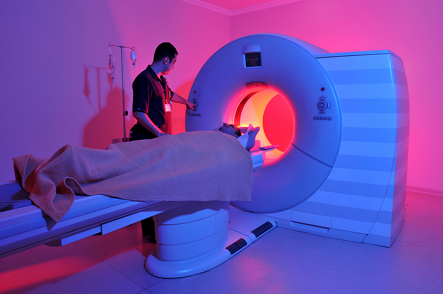
Paola Dazzan, Ph.D., and Janaina Mourao-Miranda, Ph.D., used computer algorithms to analyze magnetic resonance imaging (MRI) scans and assess a mental health patient’s outcome. The study findings are published in the journal Psychological Medicine.
Dazzan said ‘This is the first step towards being able to use brain imaging to provide tangible benefit to patients affected by psychosis.’
During psychotic periods a sufferer may suffer with hallucinations and delusions and impaired insight may also occur. Psychosis is referred to as an abnormal condition of the mind. The most common forms are associated with mental health conditions such as schizophrenia and bipolar disorder. The symptoms of psychosis can also be present in conditions such as Parkinson’s disease and drug or alcohol abuse.
Psychosis can be persistent and can affect a patient’s ability to function and lead a normal life but many patients recover from psychosis with minimal symptoms.
At present, physicians are usually unable to predict a patient’s risk of future psychosis. This uncertainty compromises a patient’s counseling and the development of a suitable treatment plan.
In this study, Dazzan and colleagues worked with 100 participant patients. MRI brain scans were taken when the participants presented to clinical services with a first psychotic episode. The researchers also scanned the brains of a control group of 91 healthy individuals.
Six years later the participants were followed up and classified as having developed a continuous episode or an intermediate illness, depending on whether their symptoms remitted or persisted during this time.
The researchers then analysed scans from 29 subjects who had a continuous course of illness, the same number from patients with an episodic course. They also analysed the same number of people from the healthy control.
The scans were used to develop pattern recognition software to differentiate between the different severities of the illness. The algorithm which was applied to the scans that were collected at the first episode of psychosis, was able to distinguish between patients who then went on to develop continuous psychosis and those who went on the develop a less severe, episodic psychosis in seven out of 10 cases.
Mourao-Miranda said ‘Although we have some way to go to improve the accuracy of these tests and validate the results on independent large samples, we have shown that in principle it should be possible to use brain scans to identify at the first episode of illness both patients who are likely to go on to have a continuous psychotic illness and those who will develop a less severe form of the illness.’
‘This suggests that even by the time that they have their first episode of psychosis, significant changes have already occurred to their brains.’
Dazzan added ‘This could, in future, offer a fast and reliable way of predicting the outcome for an individual patient allowing us to optimise treatment for those most in need, while avoiding long-term exposure to antipsychotic medications in those with very mild forms.’
‘Structural MRI scans can be obtained in as little as 10 minutes and so this technique could be incorporated into routine clinical investigations. The information this provides could help inform the treatment options available to each patient, and help us better manage their illness,’ Dazzan concluded.
Source: Wellcome Trust


